


DENTAL CARE
 We have always made dentistry a priority for our patients. Our veterinary team is dedicated to excellent oral health. One of the most important parts of our dental procedures is our ability to take digital dental radiographs. Since half of the tooth lies beneath the gum line (gingiva), the best way to evaluate the tooth and to look for underlying issues such as root resorption, fractures, or periodontal disease is via x-ray. A tooth's crown may look perfect on a physical examination of the mouth, but once the pet is anesthetized, digital dental radiographs can reveal any bone loss surrounding the tooth and root. With our feline patients, we often observe problems like tooth root absorption, an issue that is best identified via x-ray. These radiographic findings guide us as to which teeth are healthy, which teeth are salvageable, and which teeth need to be extracted. Once the diseased tooth is extracted and painful lesions addressed, pets feel happier and can eat comfortably.
X-RAY
With the help of radiography we are able to portray inner organs. In contrast to photography the image of the object x-rayed is dependant on how compact the structure of the organ is. The denser an organ is the less x-rays will be able to penetrate it and expose the film. Therefore structures such as bone, minerals (stone) or metal will show as nearly white in the final x-ray photograph.
Softer tissues will image in different shades of gray whereas air will provide a black image.
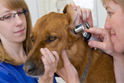
We work with a low-radiation, high frequency x-ray apparatus. We also have a specialized digital dental X-Ray device to our disposal wich enables us to perform special diagnosis of the teeth and tooth disorders.
Radiography will for instance help us to diagnose:
- Diseases of the bones (fractures, arthroses)
- Diseases of the heart and lungs (size of heart, congestion of the lungs, tumors)
- Diseases of the gastrointestinal system (constipation, flatulence, tumors, foreign bodies)
- Bladder stones
Not all structures can be shown on an x-ray photograph. Swallowed rubber or plastic balls for instants can not be differentiated from the structure around them.
SURGERY
 We perform surgery in the mornings. Should your animal need an operation we ask you to make an appointment.
We are equipped to do most kinds of soft tissue surgery. Our resources to monitor the anesthesia include Pulsoxymetrie, Kapnographie, ECG and a constant measurement of body temperature to ensure the safty of our animal-patients.
Among other things modern devices of dental medicine are at our disposal with which we are able to treat most dental disorders occurring in cats, dogs, as well as in smaller animals.
In this practice we don’t do bone surgery. Should such an operation become necessary for your animal we will be glad to councel you as to which specialized clinic you should turn to and help you make an appointment.
LAB
Through the imaging techniques we will get a picture of the specific organ. Whether or not the organ is functioning as it should, can only be determined through blood analysis.
We work with an instrument for blood analysis, which can give us the most important information within minutes.
A blood analysis can among other things inform us, whether your pet is battling an infection, whether the liver and the kidneys are functioning well or whether there are other failures in the metabolism. Furthermore hormonal disorders such as diabetes or thyroid disorders can be diagnosed this way.
For large scale laboratory examinations we cooperate with a professional lab. The blood samples will be picked up by a courier the same day. The results will then be available the next day.
ECG (Elektrocardiography)
During the different phases of the heartbeat the electrical potential in the heart muscle cell changes. The ECG measures these changes through electrodes that are attached to the animal’s skin and reproduces them graphically. For this purpose the animal must be held shortly in a recumbent position.
During the recording the electrical torrents of the heart muscle fibers are measured by electrodes that are attached to the skin by clamps.
The ECG is an important technique for determining arrhythmia as well as for diagnosing and treating other cardiac diseases.
ULTRASOUND
The technique of ultrasonic imaging is based on sound waves of a certain frequency. The sound waves are sent out by the machine and are reflected by the organs. The reflected sound waves are read and the machine produces an image of the organ.
This enables us to look into the organ. Unfortunately the ultrasound beam cannot penetrate bone, air and ingested food, so that this method is restricted to compact organs or those filled with fluids (for example liver, kidneys, spleen, heart, bladder, prostate).
ECHOCARDIOGRAPHY (Ultrasonography of the heart):
Through echocardiography the movements of the heart muscle can be evaluated.This is especially important to estimate the degree of a heart's insufficiency.
Inside view of the heart in an echocardiographic image is possible, as well as analysis of the function of the heart muscle and measurement of the ventricle as well as the heart muscle.
On the x-ray we can only see the shape of the heart. The ultrasonography enables us to assess the different chambers of the heart, to measure the thickness of the muscles and to perceive the function of the cardiac valves.
Since heart and lungs form a functional unit and the lungs can not be imaged by ultrasonography, an examination of the heart always must include an x-ray.